Media Center
A multimedia mosaic of moments at GIST
GIST Excellence
[Press Release] Upconverting nanoparticles (UCNP) used for 3D imaging of living cells
- 엘리스 리
- REG_DATE : 2016.01.07
- HIT : 895
Upconverting nanoparticles (UCNP) used for 3D imaging of living cells
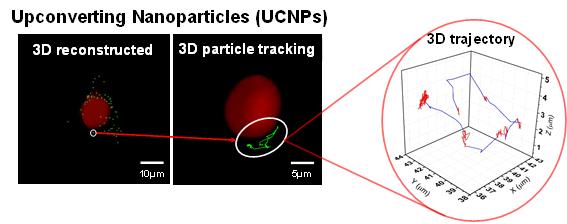
(Figure 1). The left image shows a 3D distribution of upconverting nanoparticles in green with the cell
nucleus in red, and the image on the right shows the 3D trajectory of the upconverting nanoparticles.
Most biological samples are three-dimensional (3D); however, optical microscopy techniques usually result in two-dimensional (2D) images because the focal plane of the specimen is determined by the microscope"s objective length, which creates a limited cross-section for visualization purposes. 3D images can be created using confocal or two-photon microscopy, but these techniques have limitations with imaging speed. To overcome these limitations, researchers at the Gwangju Institute of Science and Technology (GIST) have developed a simple method to construct real-time 3D images of live cells through wide-field image sectioning using lanthanide-doped upconverting nanoparticles (UCNP) as the imaging probe.
GIST Professor Min-Gon Kim and Professor Kang Taek Lee of the Department of Chemistry led Master"s student Hong Li Jo, postdoctoral researcher Dr. Yo Han Sung, and GIST College senior Jinho Park as joint-first authors of a paper entitled "Fast and background-free three-dimensional (3D) live-cell imaging with lanthanide-doped upconverting nanoparticles," which was accepted on October 25, 2015, for publication in the prestigious science journal Nanoscale with an impact factor of 7.394. Also participating in the research were Ph.D. candidate Eun-Jung Jo, GIST College senior Yeongchang Goh, and joint-degree student Kyujin Shin.
UCNP have widely been considered the most promising bioimaging probe because of several inherent advantages. For example, certain nanocrystals used for UCNP absorb 980 nm photons and emit visible photons at 525, 540, and 655 nm. Furthermore, cytotoxicity is very low while near-infrared excitation minimizes autofluorescence from and photo-damage to the cells. These characteristics combine to give UCNP discernable advantages over other imaging techniques when considering imaging speed, duration, spatiotemporal resolution, and photo-damage. Thus, UCNP can provide researchers with a powerful platform from which to study the complex intricacies of 3D cellular dynamics that have until now been obscured by the limitations of 2D imaging techniques.

(Clockwise from top left): Professor Kang Taek Lee, Professor Min-Gon Kim,
Jinho Park, Hong Li Jo, and Dr. Yo Han Song.
On that note, the paper"s co-lead research Professor Kang Taek Lee noted: "The results of this research to create real-time imaging of 3D cellular activity within living cells has the potential to revolutionize our understanding of genomic drug delivery, cancer treatment, and gene therapy as well as other critical biological processes."