Media Center
A multimedia mosaic of moments at GIST
GIST Excellence
[Press Release] APRI Dr. Joon Heon Kim: microsphere film structures can be investigated with a simple microscopy modifications
- 엘리스 리
- REG_DATE : 2015.06.12
- HIT : 858
“Easier quality control of microsphere films
with a simple microscopy modification”
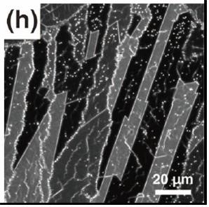
(Left) Dr. Joon Heon Kim of the Advanced Photonics Research Institute (APRI). (Fig) Image of the double-layer film consisting of various sizes of microspheres that are identifiable by their crystalline orientation.
Korean researchers have developed a method to easily identify double-layer microsphere film that are used as templates for nanostructures by using a dark-field (DF). This will enable easier and inexpensive quality control of microsphere films that are used for creating high quality nanostructures.
* Dark-field microscopy (DF) gathers lights that only bounce off a surface, making it visible with dark background. It has better contrast compared to the common bright-field microscopy (BF).
* Microsphere is a small spherical particle with the size of a couple hundred nanometers to a couple micrometers.
The research was led and performed by Dr. Joon Heon Kim of APRI. Researcher Jung-Soo Park prepared the samples and analyzed the images. Their work has been published in the online version of the Scientific Reports on May 11, 2015.
Consistence of crystalline orientation of microsphere films are considered a high-quality templates that can be used to produce high-quality nanostructures. Microscopy is used to check every microsphere’s crystalline orientation, but it takes excessive effort in identifying more sophisticated double-layer microsphere films that are used for making complex nanostructures.
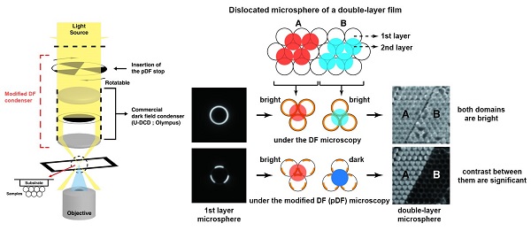
A slight modification of dark-field (DF) optical microscopy and partial dark-field (pDF) mode is a very effective technique for the visualization of domain boundaries and dislocations in close-packed double-layer microsphere films. Compared with other optical microscopic modes, this mode yields very high contrast between even very similar crystalline domains that have the same two-dimensional crystalline orientation but are slightly dislocated from each other. These domains appear as the brightest and darkest regions in each pDF image at a specific orientation of the pDF stop. By rotating the pDF stop, we can continuously change the brightness of the domains depending on their crystalline orientation. The simplicity of switching from the common full DF to the pDF mode simply with the addition of the patterned stop is also advantageous for practical applications.
Dr. Kim said, “We believe that the suggested pDF technique will extend the versatility of optical DF microscopy in the study of close-packed microsphere films, and the technique can be used in related research and in nanostructure production.
This research was supported by the Basic Science Research Program through the National Research Foundation of Korea and funded by the Ministry of Education and by the APRI Research Program through a grant provided by the Gwangju Institute of Science and Technology.