Media Center
A multimedia mosaic of moments at GIST
GIST Excellence
A Neurotransmitter Analysis System to Identify Alternatives to Botox
- 김슬혜
- REG_DATE : 2014.06.20
- HIT : 1268
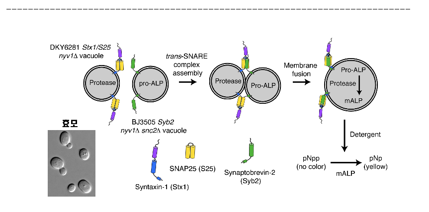
A Neurotransmitter Analysis System to Identify Alternatives to Botox - Yeast-based human nerve cell signaling mimicry helps better understand neurotransmitters - Professor Youngsoo Jun’s team published the results on PNAS □ A Korean research team has developed a system to analyze synaptic vesicle fusion*, signaling process among human nerve cells, more stably. The results are expected not only to help better understand neurotransmission processes but also to be used as a key tool to identify neurotransmitters such as Botox**. * Synaptic vesicle fusion: The process of signaling between nerve cells by which synaptic vesicles containing neurotransmitters that are mediators of brain activities such as emotions, behaviors and memory secrete neurotransmitters through nerve cell membrane fusion. ** Botox: Neurotoxin secreted by botulinum. It inhibits neurosignal transmission by eliminating SNARE protein that is involved in synaptic vesicle fusion. The substance is used in pain clinics and cosmetic treatments but has risk for side effects such as muscular paralysis and death. o Led by professor Yongsoo Jun (corresponding author) and Youngjoon Ko and Miriam Lee(co-first authors) in the School of Life Sciences at the Gwangju Institute of Science and Technology (GIST), this study was funded by the Medical Research Center Support Project of the Ministry of Science, ICT and Future Planning and the Korea Research Foundation, and the GIST Bio-imaging Research Center, and the results were published in an online edition of the Proceedings of the National Academy of Sciences (PNAS) on May 12 2014 (title: In vitro assay using engineered yeast vacuoles for neuronal SNARE-mediated membrane fusion). □ Key to signaling among nerve cells is the inside of nerve cells: how synaptic vesicles that are the container of neurotransmitters fuse with nerve cell membranes. o Using nerve cell biomembranes is the best way to analyze synaptic vesicle fusion, but it is costly and complicated. Accordingly using synthetic liposome remains the mainstream, which is easy to interpret test results but has limitations from using the synthetic material. □ Professor Jun’s research team developed yeast with human SNARE* gene and realized an in vitro model of membrane fusion between yeast** vacuoles*** to mimic human synaptic vesicle fusion. * SNARE: A protein that mediates the transport of substances in cells by inducing biomembrane fusion. ** Yeast: A monad used in bread and beer fermentation. Frequently used in in vitro studies as yeast has eucaryotic cells that have nucleus inside like human cells. *** Vacuole: A yeast cell organelle corresponding to lysosome in human cells. Decomposition and reuse of various bio-polymers such as proteins and fat occurs here. o This model overcomes the problem of reproducibility in the existing analysis systems in that vacuoles that have the same SNARE protein can be separated and refined from yeast. As yeast can be cultured in quantity, the model can be applied to large-scale screening studies to identify compounds, etc. o The study is expected to be applicable to the identification, development, and verification of compounds to mediate neurotransmission processes. □ Vacuole membrane fusion in yeast was inhibited by Botox, which is known to inhibit synaptic vesicle fusion in human nerve cells. o “The analysis system represents the characteristics of human synaptic vesicle fusion, so our study will contribute to various further studies such as improving the efficacy of Botox and developing alternatives,” said Professor Jun. Figure 1. Illustration of the synaptic vesicle fusion analysis system using engineered yeast vacuole (Left) Yeast vacuole (gray sphere marked ‘pretease’) engineered to express Syntaxin (purple) and SNAP25 (yellow) contains a protein decomposition enzyme (protease), and yeast vacuole (gray sphere marked ‘pro-ALP’) expressing Synaptobrevin (green) contains pro-alkaline phosphatase (pro-ALP). (Middle) Trans-SNARE complex between Syntaxin/SNAP25 and Synaptobrevin is formed by mixing the vacuoles together and (right) leads to membrane fusion. (Bottom) Membrane fusion results in the mix up of the contents of the two vacuoles, and protease activates pro-ALP. Quantitative measurement of membrane fusion is feasible by measuring the level of change of substrate (pNpp, colorless) to pNp (yellow) by the activated enzyme. Figure 2. Vacuole membrane fusion restrained by Botox-mediated synaptic SNARE 
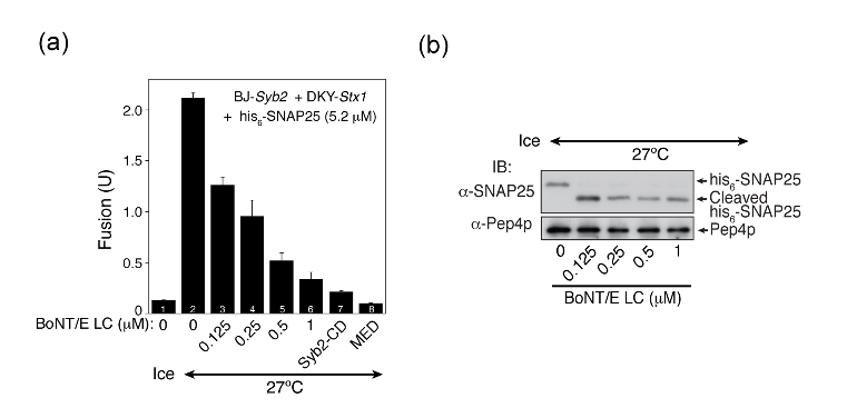
(a) Botox, a synaptic vesicle fusion inhibitor, inhibits vacuole membrane fusion in proportion to its concentration. Membrane fusion did not occur in lower temperature (Ice) but a certain level of membrane fusion was observed at 27 °C. But the membrane fusion was inhibited by increasing the concentration of Botox in pro rata basis. Membrane fusion was prevented almost completely at 1mM. In control groups, cytoplasmic domain of Synaptobrevin and phosphatide ligand MED also inhibited membrane fusion.
(b) It was observed that Botox-mediated membrane fusion inhibition was attributable to the decomposition of synaptic SNARE (SNAP25). The decomposition of SNAP25 was confirmed by immunoblotting after separating the whole reactant of membrane fusion by protein electrophoresis assay. It was observed that SNAP25 was cut and shortened by added Botox. The Pep4p protein was used as a control to show similar amounts of protein existed in all samples.